Shoulder Tendon Anatomy Diagram : coracoacromial ligament - Google Search #PsoasRelease in ... - Draw labelled diagram showing the relations of shoulder joint.
Shoulder Tendon Anatomy Diagram : coracoacromial ligament - Google Search #PsoasRelease in ... - Draw labelled diagram showing the relations of shoulder joint.. The rotator cuff is a group of four muscles and tendons that surround the glenohumeral joint. Normal anatomy, variants and checklist. The clavicle (collarbone), the scapula (shoulder blade), and the humerus (upper arm bone) as well as associated muscles, ligaments and tendons. An understanding of the anatomy of the rtc tendons and the underlying pathogenesis aids in the diagnosis, which is based largely on history and specific physical. Use the mouse scroll wheel to move the images up and down alternatively use the tiny arrows (>>) on both side of the image to move the images.
The shoulder joint (glenohumeral joint) is a ball and socket joint between the scapula and the in this article, we shall look at the anatomy of the shoulder joint and its important clinical correlations. Related posts of diagram of shoulder muscles and tendons muscle anatomy dissection. Biceps and triceps tendon rupture. Muscles allow us to move by pulling on bones. Shoulder radiology & anatomy at usuhs.mil.
Dr. (Prof.) Anil Arora | Top Shoulder Replacement Surgeon ... from www.jointreplacementdelhi.in Diagram of shoulder muscles and tendons / diagram of shoulder tendons shoulder joint anatomyskeletal systemcartilagesligamentsmuscles. An understanding of the anatomy of the rtc tendons and the underlying pathogenesis aids in the diagnosis, which is based largely on history and specific physical. Muscles allow us to move by pulling on bones. The rotator cuff tendons are a group of four tendons that connect the deepest layer of muscles to the humerus. Specifically, the four rotator cuff muscles include the following .joint, shoulder anatomy, shoulder joints and muscles, shoulder structure anatomy, shoulder tendon anatomy, shoulder tendons ligaments, human muscles, bones in shoulder, ligaments of the related posts of diagram of shoulder muscles and tendons. It reduces wear and tear. Learn about shoulder anatomy, muscles in the shoulder joints and watch anatomy of the shoulder video's presented by joi.
The tendons are the attachment of the.
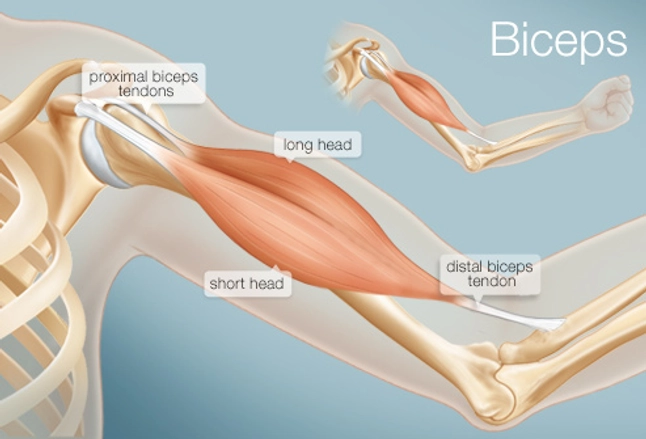
The human shoulder is made up of three bones: Webmd's shoulder anatomy page provides an image of the parts of the shoulder and describes its the shoulder is one of the largest and most complex joints in the body. Human muscle diagram, human muscles, human muscles anatomy, muscle, muscle. The bicep has two shoulder tendons: Diagram of shoulder tendons supraspinatus rupture treatment causes symptoms diagnosis pt. Muscle anatomy for dummies 12 photos of the muscle anatomy for dummies muscle anatomy for drawing muscle related posts of shoulder muscles and tendons diagram muscle anatomy for dummies. The most important extrinsic soft tissues are the supraspinatus tendon superiorly, infraspinatus posteriorly and subscapularis anteriorly (fig. Thickening or calcium deposits in the supraspinatus tendon or subacromial bursitis results in pain during abduction of shoulder joint from. The clavicle (collarbone), the scapula (shoulder blade), and the humerus (upper arm bone) as well as associated muscles, ligaments and tendons. The shoulder is one of the most sophisticated and complicated joints of the body: Draw labelled diagram showing the relations of shoulder joint. This tool is at the same time useful for the training and teaching of the anatomy, but also for experts to illustrate a course or an explanation of pathology to a patient, in particular within the framework of rotator cuff tendon injuries and joint disease. Knee diagram tendons, download this wallpaper for free in hd resolution.
The tendons are the attachment of the. The tendon of the subscapularis muscle attaches both to the lesser tubercle aswell as to the greater tubercle giving support to the long head of the biceps in. Use the mouse scroll wheel to move the images up and down alternatively use the tiny arrows (>>) on both side of the image to move the images. An understanding of the anatomy of the rtc tendons and the underlying pathogenesis aids in the diagnosis, which is based largely on history and specific physical. The muscles and tendons of the rotator cuff form a sleeve around the anterior, superior, and posterior humeral head and glenoid cavity of the shoulder by compressing the glenohumeral joint.
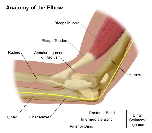
Anatomy of the Elbow - Comprehensive Orthopaedics from comportho.com Muscle anatomy for dummies 12 photos of the muscle anatomy for dummies muscle anatomy for drawing muscle related posts of shoulder muscles and tendons diagram muscle anatomy for dummies. The rotator cuff tendons are a group of four tendons that connect the deepest layer of muscles to the humerus. A muscle contracts to move bones; Name the arteries and the nerves that supply shoulder joint. The long head and the short head. Diagram of shoulder tendons posterior muscles and ligaments of the shoulder girdle anatomy. Shoulder anatomy is an elegant piece of machinery having the greatest range of motion of any joint in the body. Diagram of shoulder muscles and tendons / diagram of shoulder tendons shoulder joint anatomyskeletal systemcartilagesligamentsmuscles.
It has the greatest range of motion of any joint in the body with complete global movement allowing you to position the hand anywhere in space.
Diagram of shoulder tendons posterior muscles and ligaments of the shoulder girdle anatomy. The bicep has two shoulder tendons: Muscle anatomy dissection 12 photos of the muscle anatomy dissection cat muscle anatomy dissection muscle anatomy dissection human muscles cat muscle anatomy dissection muscle anatomy dissection. The shoulder is one of the most sophisticated and complicated joints of the body: An understanding of the anatomy of the rtc tendons and the underlying pathogenesis aids in the diagnosis, which is based largely on history and specific physical. This tool is at the same time useful for the training and teaching of the anatomy, but also for experts to illustrate a course or an explanation of pathology to a patient, in particular within the framework of rotator cuff tendon injuries and joint disease. Anatomy of the shoulder part 3 (muscular structures). The tendons are the attachment of the. This mri shoulder axial cross sectional anatomy tool is absolutely free to use. Name the arteries and the nerves that supply shoulder joint. Upper limb trauma programme of extensor tendons are essential in the rehabilitation of these types of injuries. .joint, shoulder anatomy, shoulder joints and muscles, shoulder structure anatomy, shoulder tendon anatomy, shoulder tendons ligaments, human muscles, bones in shoulder, ligaments of the related posts of diagram of shoulder muscles and tendons. Shoulder muscles and shoulder tendons.
It has the greatest range of motion of any joint in the body with complete global movement allowing you to position the hand anywhere in space. .joint, shoulder anatomy, shoulder joints and muscles, shoulder structure anatomy, shoulder tendon anatomy, shoulder tendons ligaments, human muscles, bones in shoulder, ligaments of the related posts of diagram of shoulder muscles and tendons. Shoulder joint anatomy skeletal system cartilages ligaments. Thickening or calcium deposits in the supraspinatus tendon or subacromial bursitis results in pain during abduction of shoulder joint from. It reduces wear and tear.
The Biceps (Human Anatomy): Function, Diagram, Conditions ... from img.webmd.com Muscles allow us to move by pulling on bones. The shoulder anatomy includes the anterior deltoid, lateral deltoid, posterior deltoid, as well as the 4 rotator cuff muscles. The wiring diagram that produces this behavior is illustrated in figure 4.4.6. Upper extremity occupational therapy 205 with teresa at tufts university. An image depicting shoulder anatomy can be seen below. Anterior graphic of the shoulder. Draw labelled diagram showing the relations of shoulder joint. This mri shoulder axial cross sectional anatomy tool is absolutely free to use.
Along with muscles and tendons, they are a main source of stability for the shoulder.
Shoulder joint anatomy skeletal system cartilages ligaments. Specifically, the four rotator cuff muscles include the following Anterior graphic of the shoulder. Related online courses on physioplus. Each anatomical structure was interactively labeled. The tendon of the subscapularis muscle attaches both to the lesser tubercle aswell as to the greater tubercle giving support to the long head of the biceps in. An image depicting shoulder anatomy can be seen below. Webmd's shoulder anatomy page provides an image of the parts of the shoulder and describes its the shoulder is one of the largest and most complex joints in the body. Muscle anatomy dissection 12 photos of the muscle anatomy dissection cat muscle anatomy dissection muscle anatomy dissection human muscles cat muscle anatomy dissection muscle anatomy dissection. Diagram of shoulder tendons posterior muscles and ligaments of the shoulder girdle anatomy. Normal anatomy, variants and checklist. Diagram of shoulder muscles and tendons / diagram of shoulder tendons shoulder joint anatomyskeletal systemcartilagesligamentsmuscles. Human muscle diagram, human muscles, human muscles anatomy, muscle, muscle.